Initial Examination and Dental Diagnosis

The initial examination is a critical step for a holistic evaluation of oral health. With intraoral checks, digital radiology, and early diagnosis protocols, a personalized treatment plan is prepared.

Initial Examination and Dental Diagnosis

Intraoral Examination

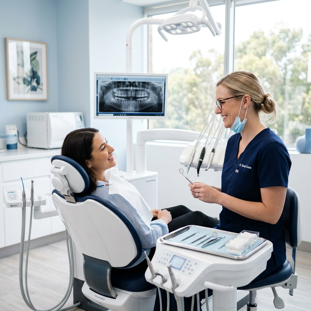
At DentARF, every patient's journey begins with a thorough intraoral examination carried out by our specialist dentists. During this comprehensive assessment, all teeth surfaces, gingival tissue, soft tissues of the mouth (cheeks, tongue, palate, floor of the mouth), and the bite relationship are carefully evaluated.

Using dental mirrors, probes, and intraoral cameras, the dentist records the condition of existing restorations, identifies carious lesions at various stages, and detects signs of gum disease such as pocket depth, bleeding on probing, and gingival recession. Every finding is documented in the patient's digital file, allowing for tracking progress across multiple visits.

For new patients, this step also includes a medical history review. Systemic conditions such as diabetes, cardiovascular disease, or immune disorders can significantly affect oral health and treatment decisions. Our clinicians review all current medications to anticipate any interactions or healing considerations.

The intraoral examination not only identifies current problems but also highlights risk factors — enabling our team to recommend preventive measures before conditions worsen. Patients receive a clear verbal explanation of all findings, and visual aids or screen displays are used to ensure full understanding.

Dental Radiology and Tomography

Modern dentistry relies on high-precision imaging to see what cannot be detected by eye alone. At DentARF, we utilize digital panoramic X-rays (OPG), periapical films, and cone-beam computed tomography (CBCT) depending on each patient's clinical need.

Digital X-rays emit up to 90% less radiation than conventional film-based systems and deliver instant, high-resolution images that can be enlarged, measured, and shared digitally. Periapical radiographs reveal root morphology, surrounding bone levels, and periapical pathology with excellent detail.

For implant planning, orthodontic assessment, or evaluation of impacted teeth and jaw pathologies, CBCT (3D tomography) provides a three-dimensional view of hard and soft tissue structures. This allows precise measurement of bone volume, identification of anatomical landmarks such as the inferior alveolar nerve and maxillary sinus, and accurate surgical simulation before any procedure.

All radiographic data is stored securely in our practice management system and can be forwarded to referring specialists when multidisciplinary care is required. Imaging is performed only when clinically justified, in line with the ALARA (As Low As Reasonably Achievable) radiation safety principle.

Risk Assessment and Treatment Planning

Following the clinical examination and radiographic review, our dentist conducts a structured risk assessment. Caries risk, periodontal risk, occlusal risk, and aesthetic concerns are each evaluated individually. This multi-axis approach ensures that no aspect of oral health is overlooked.

Based on this assessment, a phased and prioritized treatment plan is created. Acute problems (pain, infection, trauma) are addressed first. This is followed by disease control (caries removal, periodontal therapy), functional rehabilitation (restorations, prosthetics, implants), and finally aesthetic enhancement if desired.

Every plan is tailored to the patient's age, expectations, systemic health, and budget. At DentARF, we believe informed patients make better decisions — so we take the time to walk through each proposed treatment, explain the alternatives, and answer every question before any consent is given.

Early Diagnosis: The Key to Simpler Treatment

The single greatest factor in keeping dental treatment simple, affordable, and comfortable is early detection. A cavity diagnosed at the enamel stage requires only a small composite filling. The same cavity, left untreated, may progress to the pulp and require root canal therapy, a crown, or even extraction.

At DentARF, we use fluorescence-assisted caries detection and intraoral cameras in addition to standard examination techniques. These tools reveal demineralization and early carious lesions that are invisible to conventional probing or visual inspection.

Similarly, early-stage periodontal disease (gingivitis) is fully reversible with professional cleaning and improved home care. Periodontitis, its advanced form involving bone loss, requires far more extensive intervention. Regular check-up appointments every six months are our strongest recommendation for maintaining long-term oral health at minimal cost and effort.

DentARF Klinik

Take the First Step for a Healthier Smile Today

Use our online booking flow to create your appointment in 5 quick steps.

Book Appointment